Patents Directory for Dr. Gary K. Michelson — U.S. Published Patents only
340 patents found - Page 15
| Patent | Abstract |
|---|---|
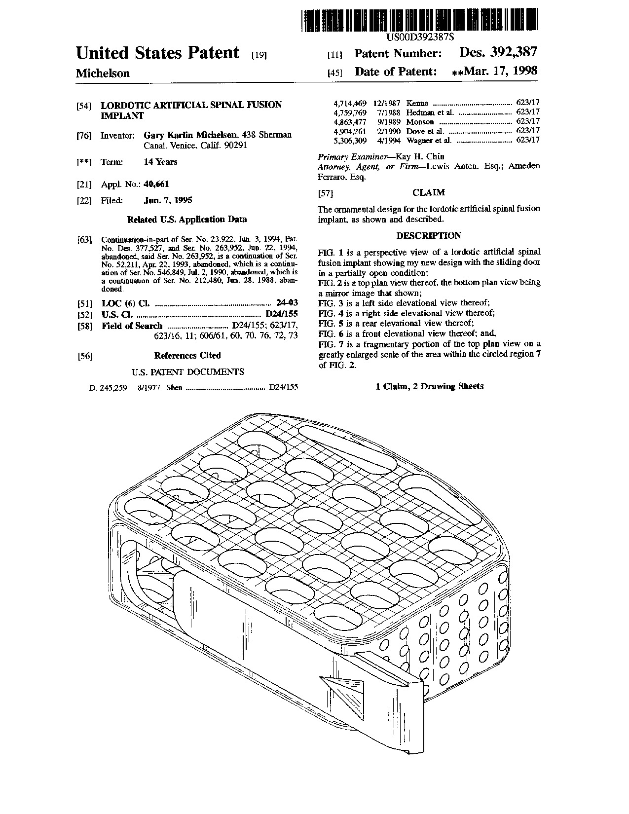 Lordotic artificial spinal fusion implant - Patent D392,387 Lordotic artificial spinal fusion implant - Patent D392,387
|
|
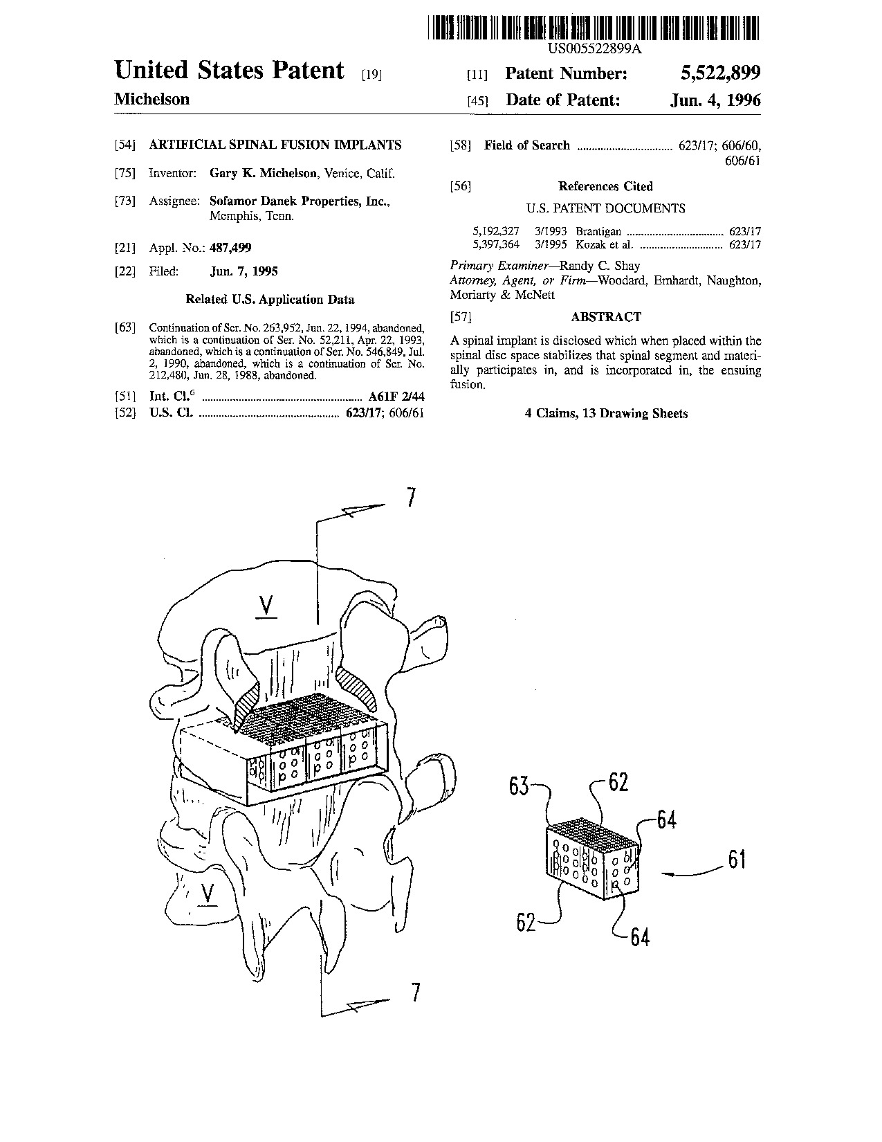 Artificial spinal fusion implants - Patent 5,522,899 Artificial spinal fusion implants - Patent 5,522,899
|
A spinal implant is disclosed which when placed within the spinal disc space stabilizes that spinal segment and materially participates in, and is incorporated in, the ensuing fusion.
|
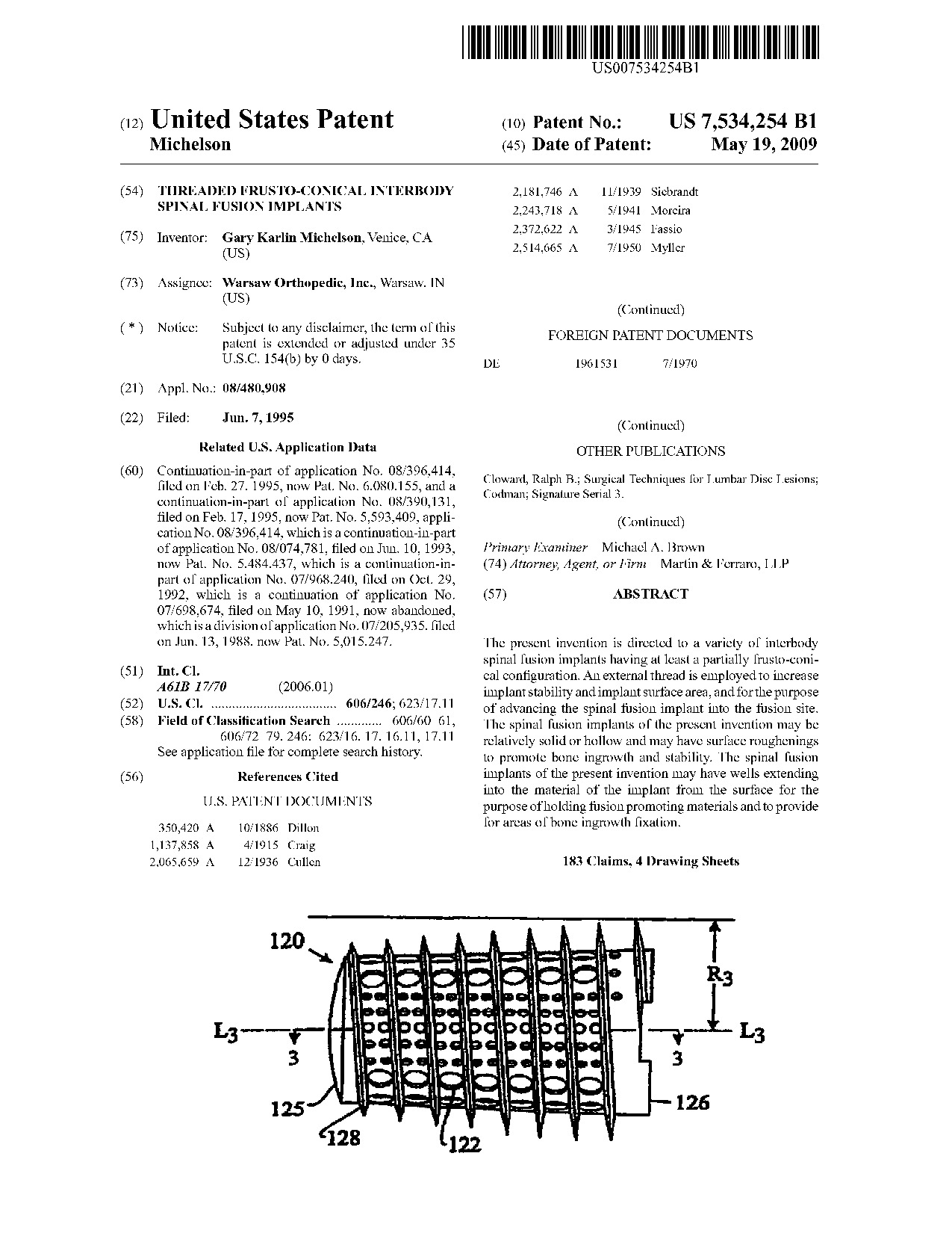 Threaded frusto-conical interbody spinal fusion implants - Patent 7,534,254 Threaded frusto-conical interbody spinal fusion implants - Patent 7,534,254
|
The present invention is directed to a variety of interbody spinal fusion implants having at least a partially frusto-conical configuration. An external thread is employed to increase implant stability and implant surface area, and for the purpose of advancing the spinal fusion implant into the fusion site. The spinal fusion implants of the present invention may be relatively solid or hollow and may have surface roughenings to promote bone ingrowth and stability. The spinal fusion implants of the present invention may have wells extending into the material of the implant from the surface for the purpose of holding fusion promoting materials and to provide for areas of bone ingrowth fixation.
|
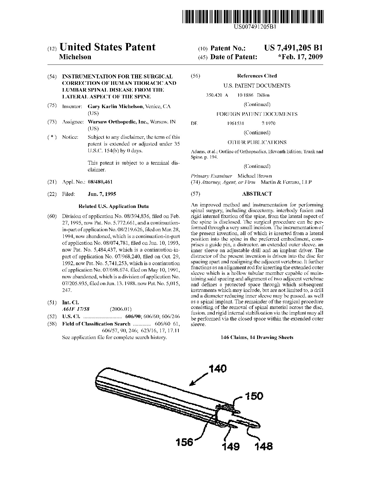 Instrumentation for the surgical correction of human thoracic and lumbar spinal disease from the lateral aspect of the spine - Patent 7,491,205 Instrumentation for the surgical correction of human thoracic and lumbar spinal disease from the lateral aspect of the spine - Patent 7,491,205
|
An improved method and instrumentation for performing spinal surgery, including discectomy, interbody fusion and rigid internal fixation of the spine, from the lateral aspect of the spine is disclosed. The surgical procedure can be performed through a very small incision. The instrumentation of the present invention, all of which is inserted from a lateral position into the spine in the preferred embodiment, comprises a guide pin, a distractor, an extended outer sleeve, an inner sleeve an adjustable drill and an implant driver. The distractor of the present invention is driven into the disc for spacing apart and realigning the adjacent vertebrae. It further functions as an alignment rod for inserting the extended outer sleeve which is a hollow tubular member capable of maintaining said spacing and alignment of two adjacent vertebrae and defines a protected space through which subsequent instruments which may include, but are not limited to, a drill and a diameter reducing inner sleeve may be passed, as well as a spinal implant. The remainder of the surgical procedure consisting of the removal of spinal material across the disc, fusion, and rigid internal stabilization via the implant may all be performed via the closed space within the extended outer sleeve.
|
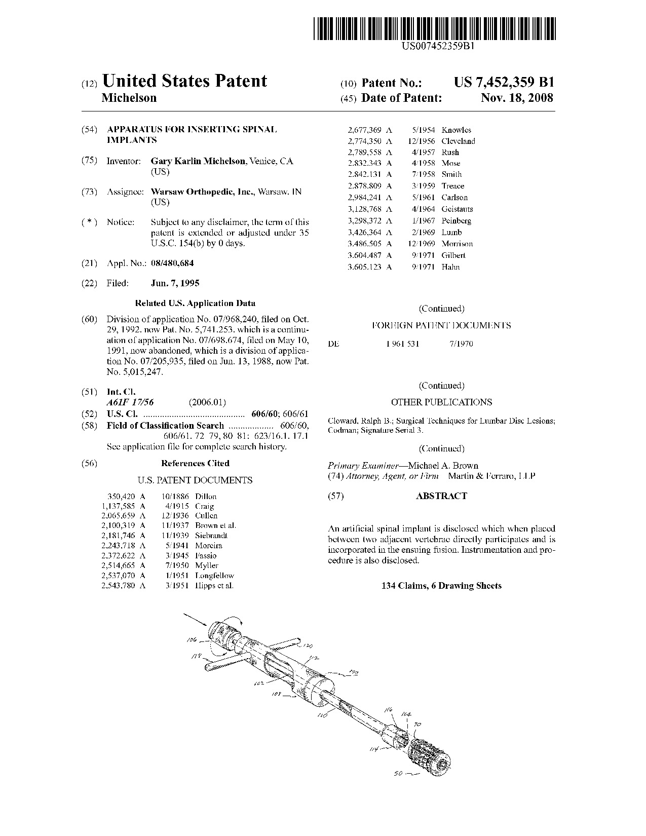 Apparatus for inserting spinal implants - Patent 7,452,359 Apparatus for inserting spinal implants - Patent 7,452,359
|
An artificial spinal implant is disclosed which when placed between two adjacent vertebrae directly participates and is incorporated in the ensuing fusion. Instrumentation and procedure is also disclosed.
|
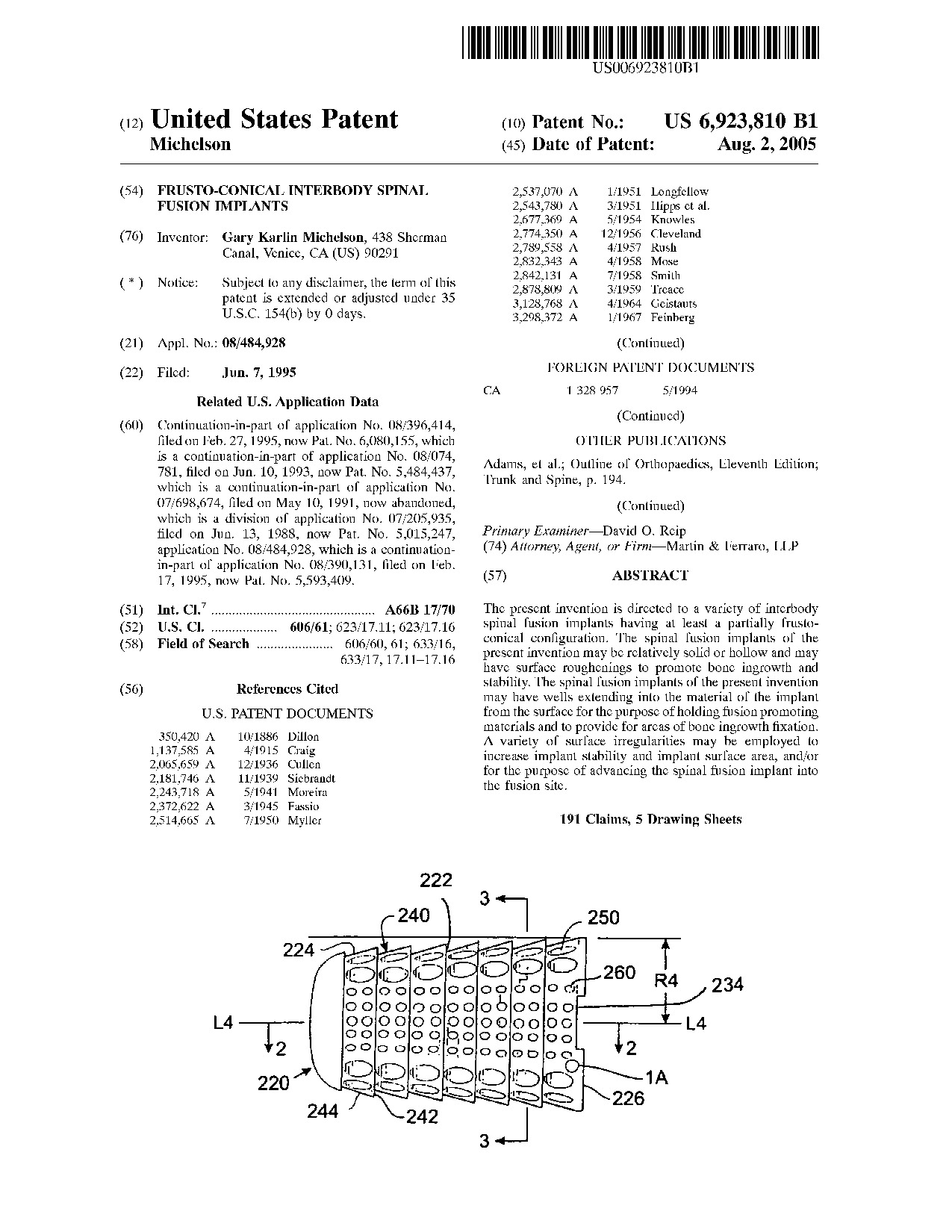 Frusto-conical interbody spinal fusion implants - Patent 6,923,810 Frusto-conical interbody spinal fusion implants - Patent 6,923,810
|
The present invention is directed to a variety of interbody spinal fusion implants having at least a partially frusto-conical configuration. The spinal fusion implants of the present invention may be relatively solid or hollow and may have surface roughenings to promote bone ingrowth and stability. The spinal fusion implants of the present invention may have wells extending into the material of the implant from the surface for the purpose of holding fusion promoting materials and to provide for areas of bone ingrowth fixation. A variety of surface irregularities may be employed to increase implant stability and implant surface area, and/or for the purpose of advancing the spinal fusion implant into the fusion site.
|
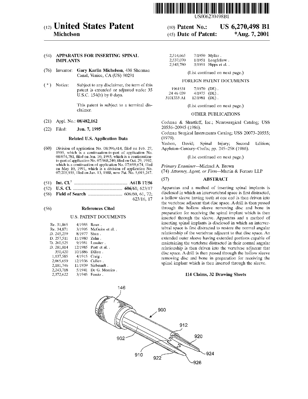 Apparatus for inserting spinal implants - Patent 6,270,498 Apparatus for inserting spinal implants - Patent 6,270,498
|
Apparatus and a method of inserting spinal implants is disclosed in which an intervertebral space is first distracted, a hollow sleeve having teeth at one end is then driven into the vertebrae adjacent that disc space. A drill is then passed through the hollow sleeve removing disc and bone in preparation for receiving the spinal implant which is then inserted through the sleeve. Apparatus and a method of inserting spinal implants is disclosed in which an intervertebral space is first distracted to restore the normal angular relationship of the vertebrae adjacent to that disc space. An extended outer sleeve having extended portions capable of maintaining the vertebrae distracted in their normal angular relationship is then driven into the vertebrae adjacent that disc space. A drill is then passed through the hollow sleeve removing disc and bone in preparation for receiving the spinal implant which is then inserted through the sleeve.
|
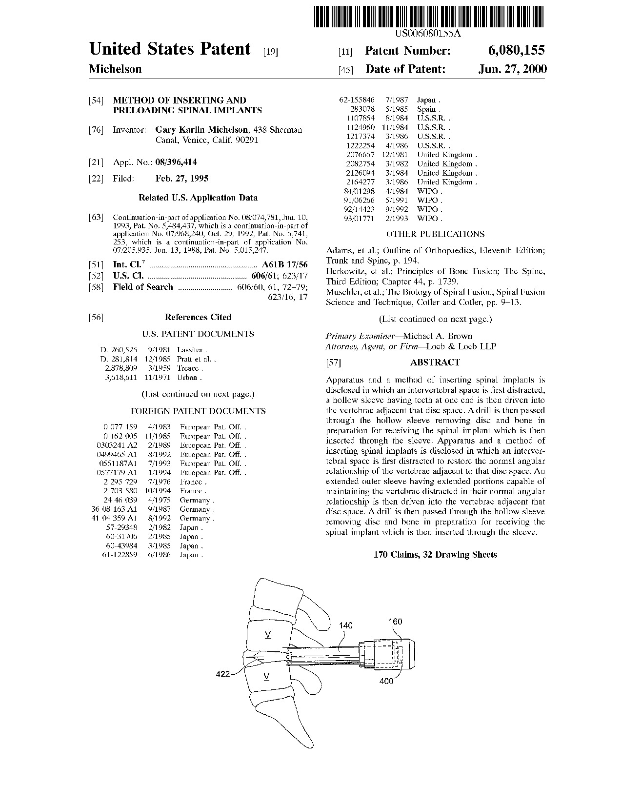 Method of inserting and preloading spinal implants - Patent 6,080,155 Method of inserting and preloading spinal implants - Patent 6,080,155
|
Apparatus and a method of inserting spinal implants is disclosed in which an intervertebral space is first distracted, a hollow sleeve having teeth at one end is then driven into the vertebrae adjacent that disc space. A drill is then passed through the hollow sleeve removing disc and bone in preparation for receiving the spinal implant which is then inserted through the sleeve. Apparatus and a method of inserting spinal implants is disclosed in which an intervertebral space is first distracted to restore the normal angular relationship of the vertebrae adjacent to that disc space. An extended outer sleeve having extended portions capable of maintaining the vertebrae distracted in their normal angular relationship is then driven into the vertebrae adjacent that disc space. A drill is then passed through the hollow sleeve removing disc and bone in preparation for receiving the spinal implant which is then inserted through the sleeve.
|
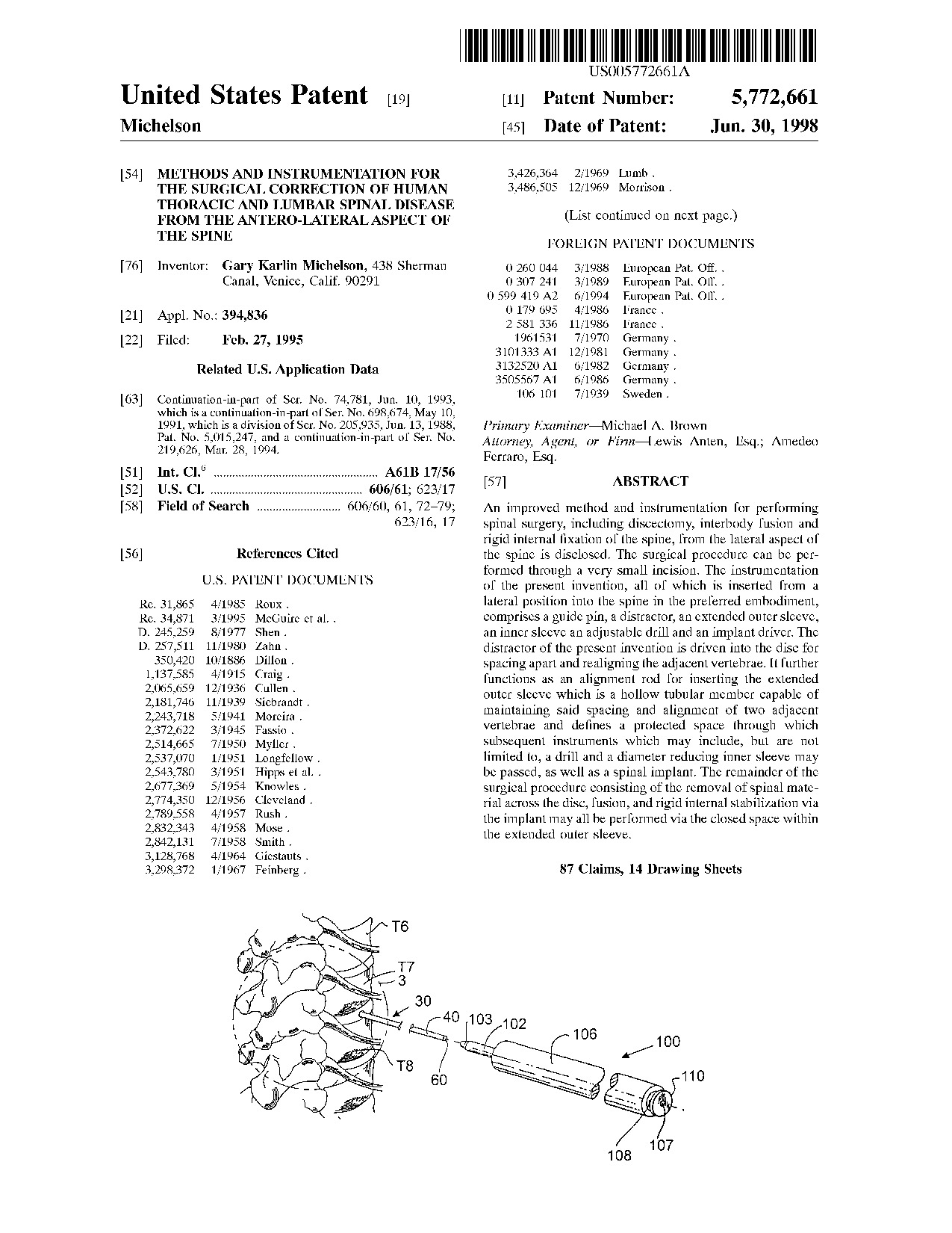 Methods and instrumentation for the surgical correction of human thoracic and lumbar spinal disease from the antero-lateral aspect of the spine - Patent 5,772,661 Methods and instrumentation for the surgical correction of human thoracic and lumbar spinal disease from the antero-lateral aspect of the spine - Patent 5,772,661
|
An improved method and instrumentation for performing spinal surgery, including discectomy, interbody fusion and rigid internal fixation of the spine, from the lateral aspect of the spine is disclosed. The surgical procedure can be performed through a very small incision. The instrumentation of the present invention, all of which is inserted from a lateral position into the spine in the preferred embodiment, comprises a guide pin, a distractor, an extended outer sleeve, an inner sleeve an adjustable drill and an implant driver. The distractor of the present invention is driven into the disc for spacing apart and realigning the adjacent vertebrae. It further functions as an alignment rod for inserting the extended outer sleeve which is a hollow tubular member capable of maintaining said spacing and alignment of two adjacent vertebrae and defines a protected space through which subsequent instruments which may include, but are not limited to, a drill and a diameter reducing inner sleeve may be passed, as well as a spinal implant. The remainder of the surgical procedure consisting of the removal of spinal material across the disc, fusion, and rigid internal stabilization via the implant may all be performed via the closed space within the extended outer sleeve.
|
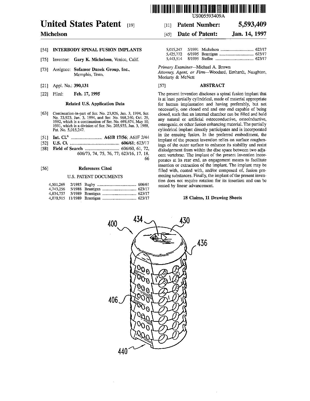 Interbody spinal fusion implants - Patent 5,593,409 Interbody spinal fusion implants - Patent 5,593,409
|
The present invention discloses a spinal fusion implant that is at least partially cylindrical, made of material appropriate for human implantation and having preferably, but not necessarily, one closed end and one end capable of being closed, such that an internal chamber can be filled and hold any natural or artificial osteoconductive, osteoinductive, osteogenic, or other fusion enhancing material. The partially cylindrical implant directly participates and is incorporated in the ensuing fusion. In the preferred embodiment, the implant of the present invention relies on surface roughenings of the outer surface to enhance its stability and resist dislodgement from within the disc space between two adjacent vertebrae. The implant of the present invention incorporates at its rear end, an engagement means to facilitate insertion or extraction of the implant. The implant may be filled with, coated with, and/or composed of, fusion promoting substances. Finally, the implant of the present invention does not require rotation for its insertion and can be seated by linear advancement.
|
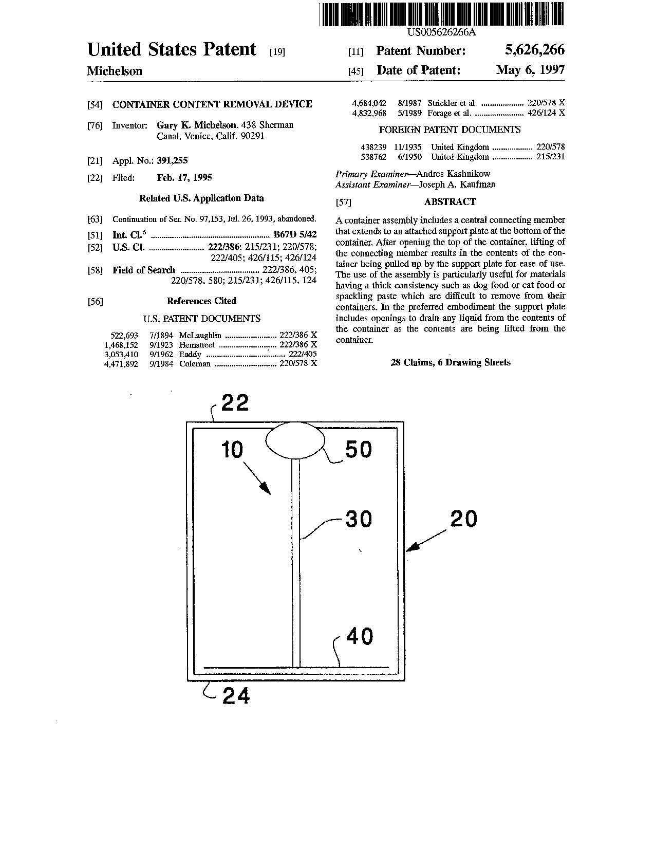 Container content removal device - Patent 5,626,266 Container content removal device - Patent 5,626,266
|
A container assembly includes a central connecting member that extends to an attached support plate at the bottom of the container. After opening the top of the container, lifting of the connecting member results in the contents of the container being pulled up by the support plate for ease of use. The use of the assembly is particularly useful for materials having a thick consistency such as dog food or cat food or spackling paste which are difficult to remove from their containers. In the preferred embodiment the support plate includes openings to drain any liquid from the contents of the container as the contents are being lifted from the container.
|
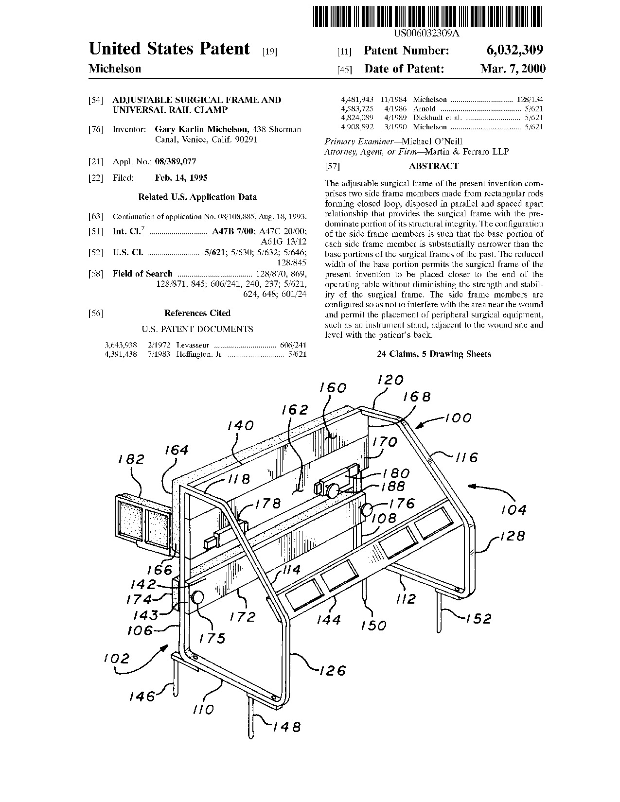 Adjustable surgical frame and universal rail clamp - Patent 6,032,309 Adjustable surgical frame and universal rail clamp - Patent 6,032,309
|
The adjustable surgical frame of the present invention comprises two side frame members made from rectangular rods forming closed loop, disposed in parallel and spaced apart relationship that provides the surgical frame with the predominate portion of its structural integrity. The configuration of the side frame members is such that the base portion of each side frame member is substantially narrower than the base portions of the surgical frames of the past. The reduced width of the base portion permits the surgical frame of the present invention to be placed closer to the end of the operating table without diminishing the strength and stability of the surgical frame. The side frame members are configured so as not to interfere with the area near the wound and permit the placement of peripheral surgical equipment, such as an instrument stand, adjacent to the wound site and level with the patient's back.
|
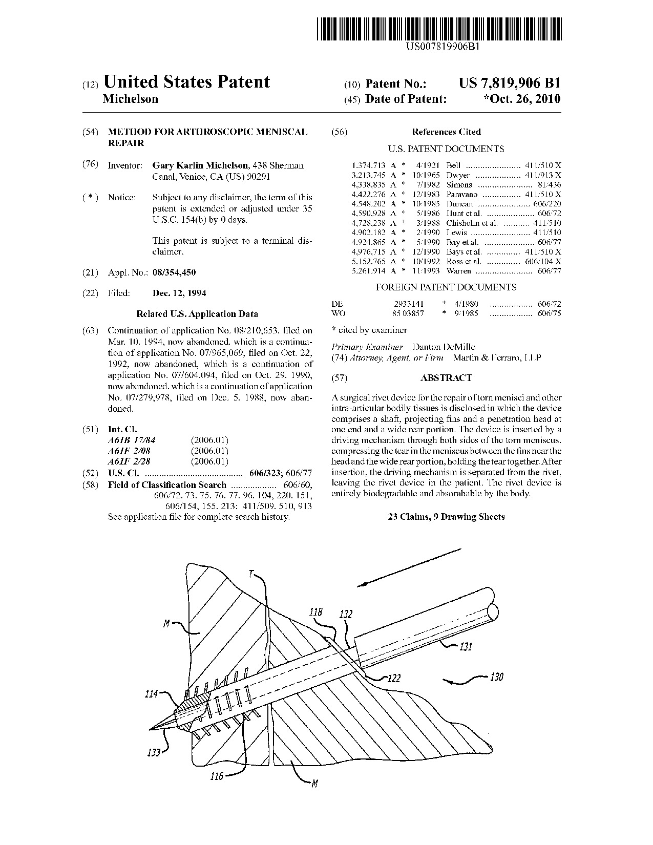 Method for arthroscopic meniscal repair - Patent 7,819,906 Method for arthroscopic meniscal repair - Patent 7,819,906
|
A surgical rivet device for the repair of torn menisci and other intra-articular bodily tissues is disclosed in which the device comprises a shaft, projecting fins and a penetration head at one end and a wide rear portion. The device is inserted by a driving mechanism through both sides of the torn meniscus, compressing the tear in the meniscus between the fins near the head and the wide rear portion, holding the tear together. After insertion, the driving mechanism is separated from the rivet, leaving the rivet device in the patient. The rivet device is entirely biodegradable and absorabable by the body.
|
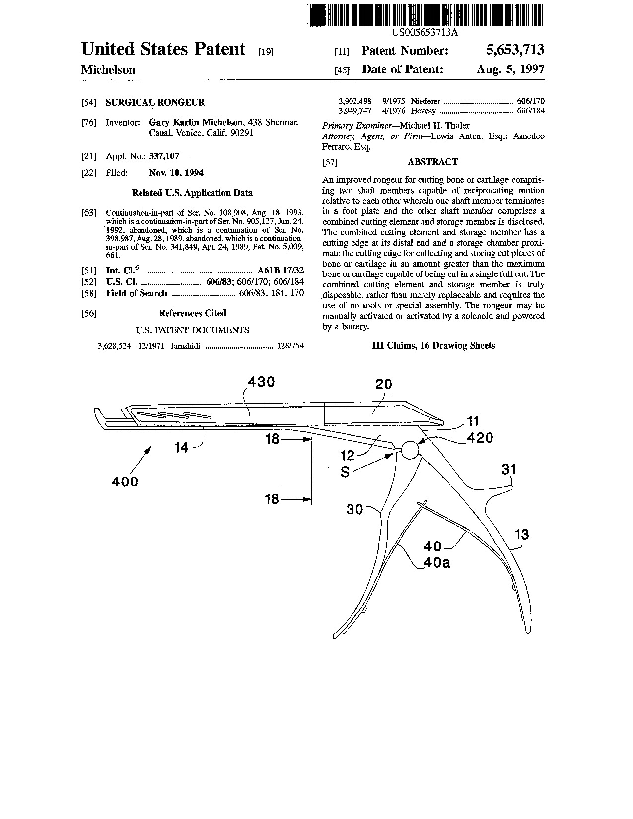 Surgical rongeur - Patent 5,653,713 Surgical rongeur - Patent 5,653,713
|
An improved rongeur for cutting bone or cartilage comprising two shaft members capable of reciprocating motion relative to each other wherein one shaft member terminates in a foot plate and the other shaft member comprises a combined cutting element and storage member is disclosed. The combined cutting element and storage member has a cutting edge at its distal end and a storage chamber proximate the cutting edge for collecting and storing cut pieces of bone or cartilage in an amount greater than the maximum bone or cartilage capable of being cut in a single full cut. The combined cutting element and storage member is truly disposable, rather than merely replaceable and requires the use of no tools or special assembly. The rongeur may be manually activated or activated by a solenoid and powered by a battery.
|
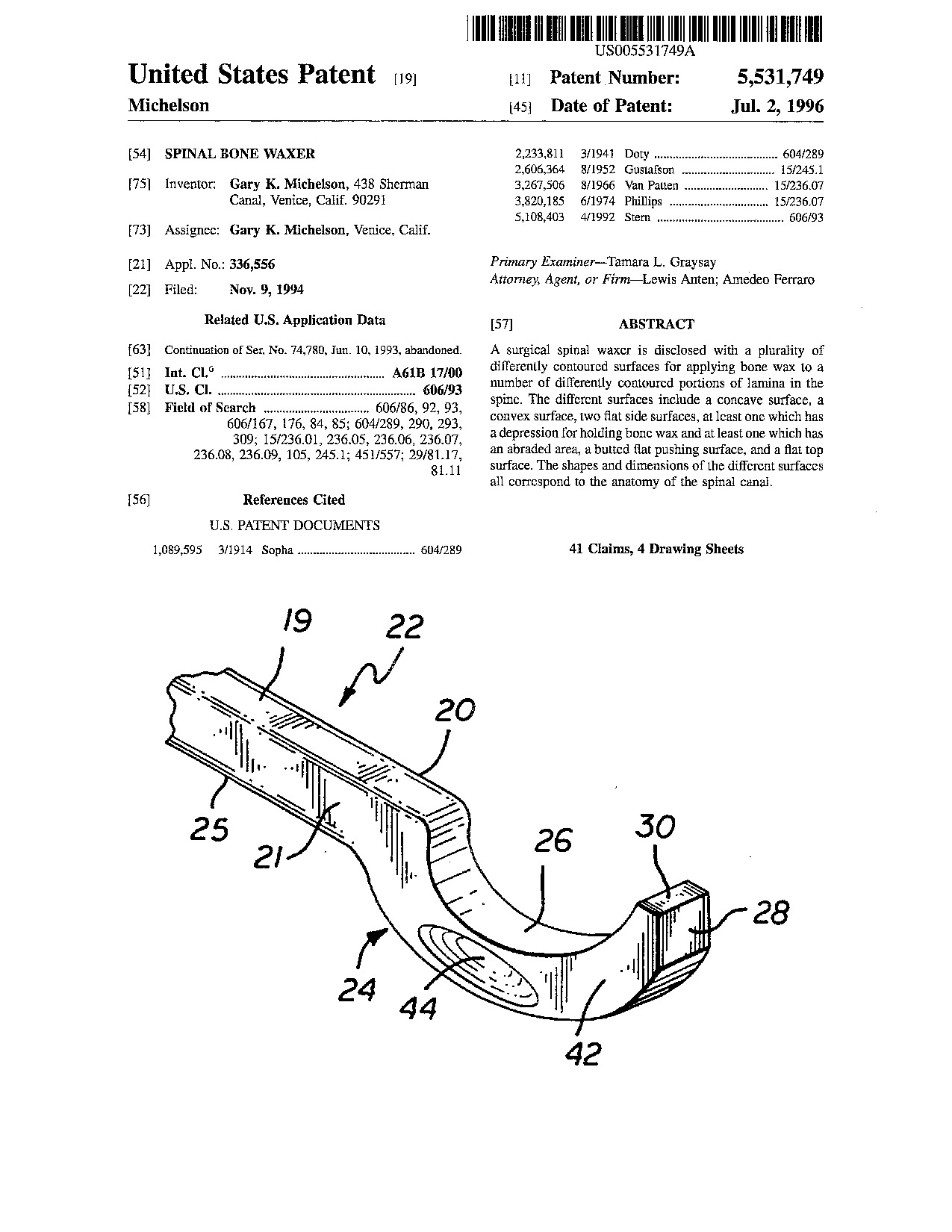 Spinal bone waxer - Patent 5,531,749 Spinal bone waxer - Patent 5,531,749
|
A surgical spinal waxer is disclosed with a plurality of differently contoured surfaces for applying bone wax to a number of differently contoured portions of lamina in the spine. The different surfaces include a concave surface, a convex surface, two flat side surfaces, at least one which has a depression for holding bone wax and at least one which has an abraded area, a butted flat pushing surface, and a flat top surface. The shapes and dimensions of the different surfaces all correspond to the anatomy of the spinal canal.
|
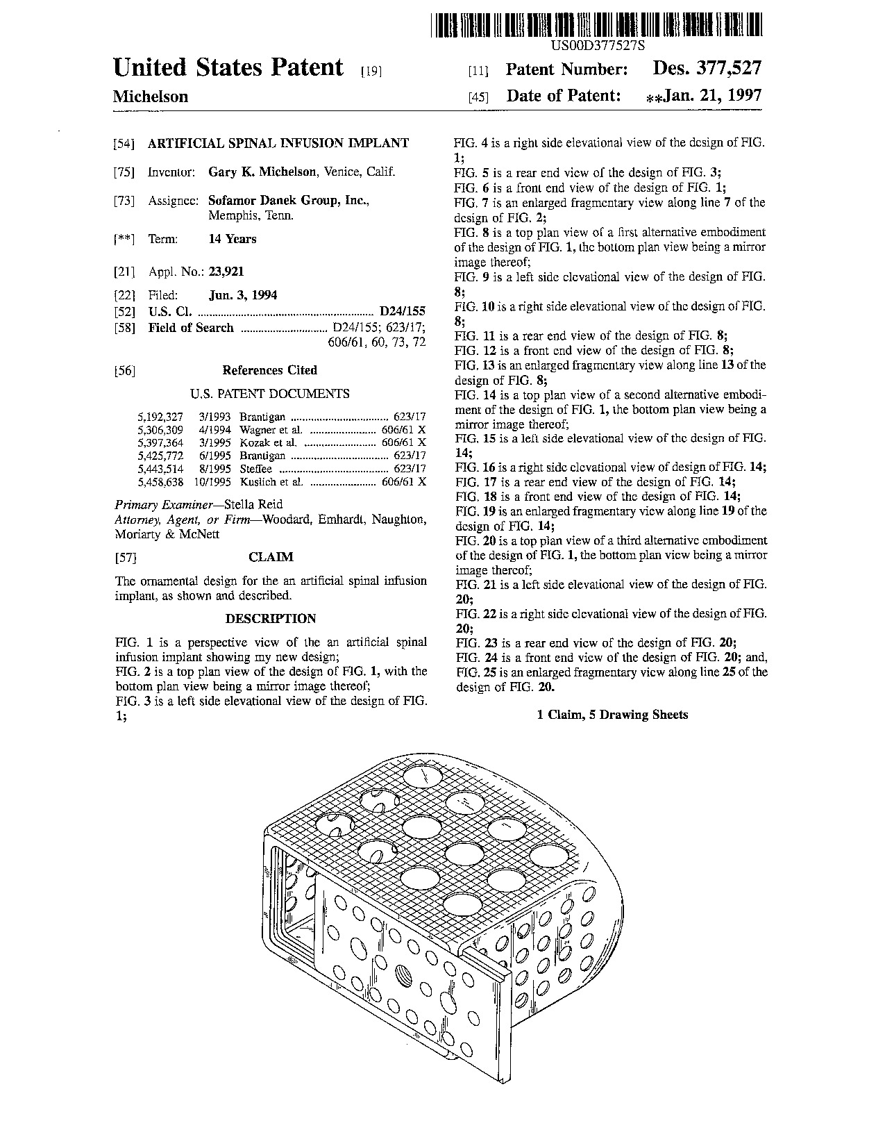 Artificial spinal infusion implant - Patent D377,527 Artificial spinal infusion implant - Patent D377,527
|
|
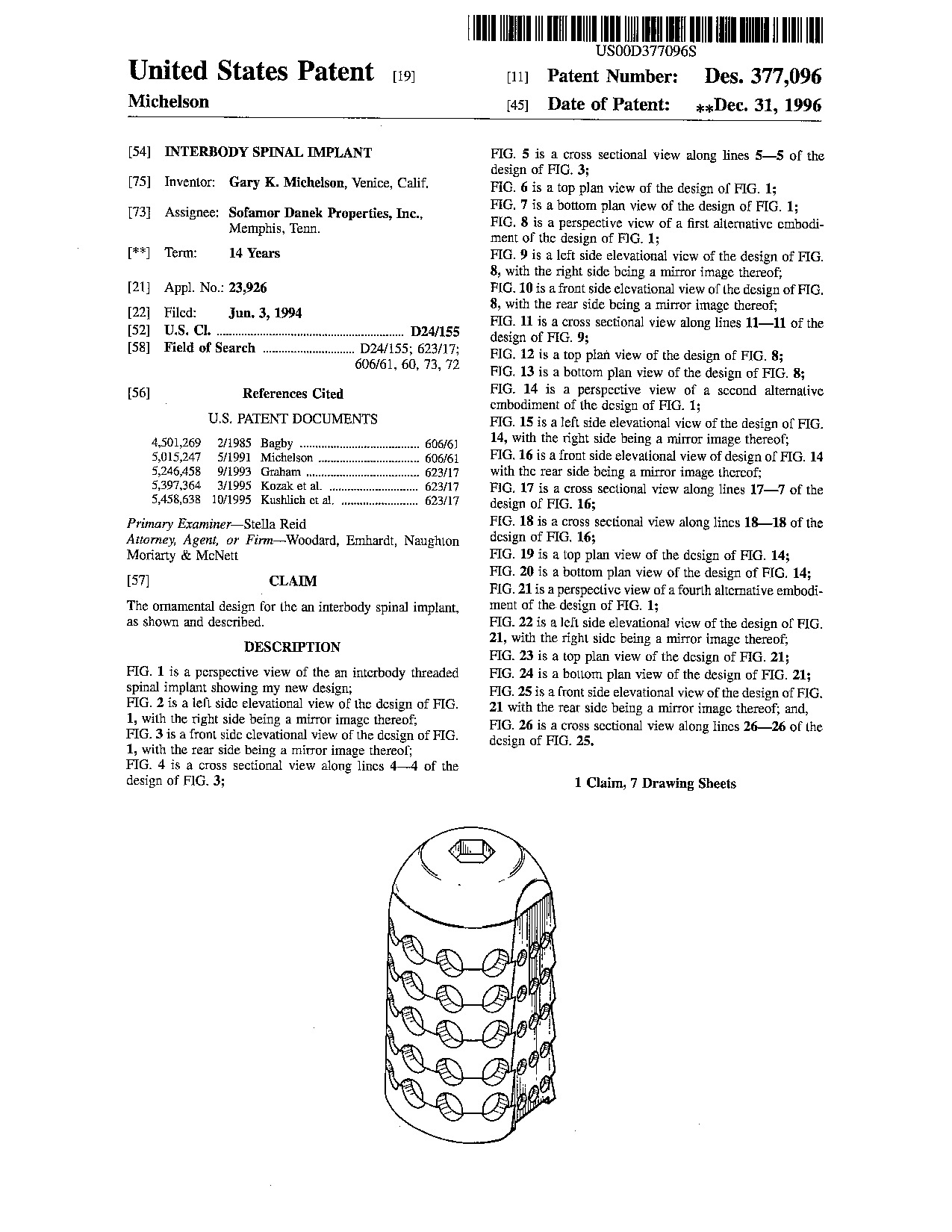 Interbody spinal implant - Patent D377,096 Interbody spinal implant - Patent D377,096
|
|
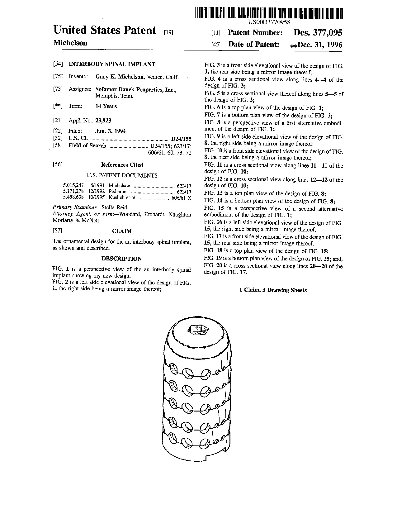 Interbody spinal implant - Patent D377,095 Interbody spinal implant - Patent D377,095
|
|
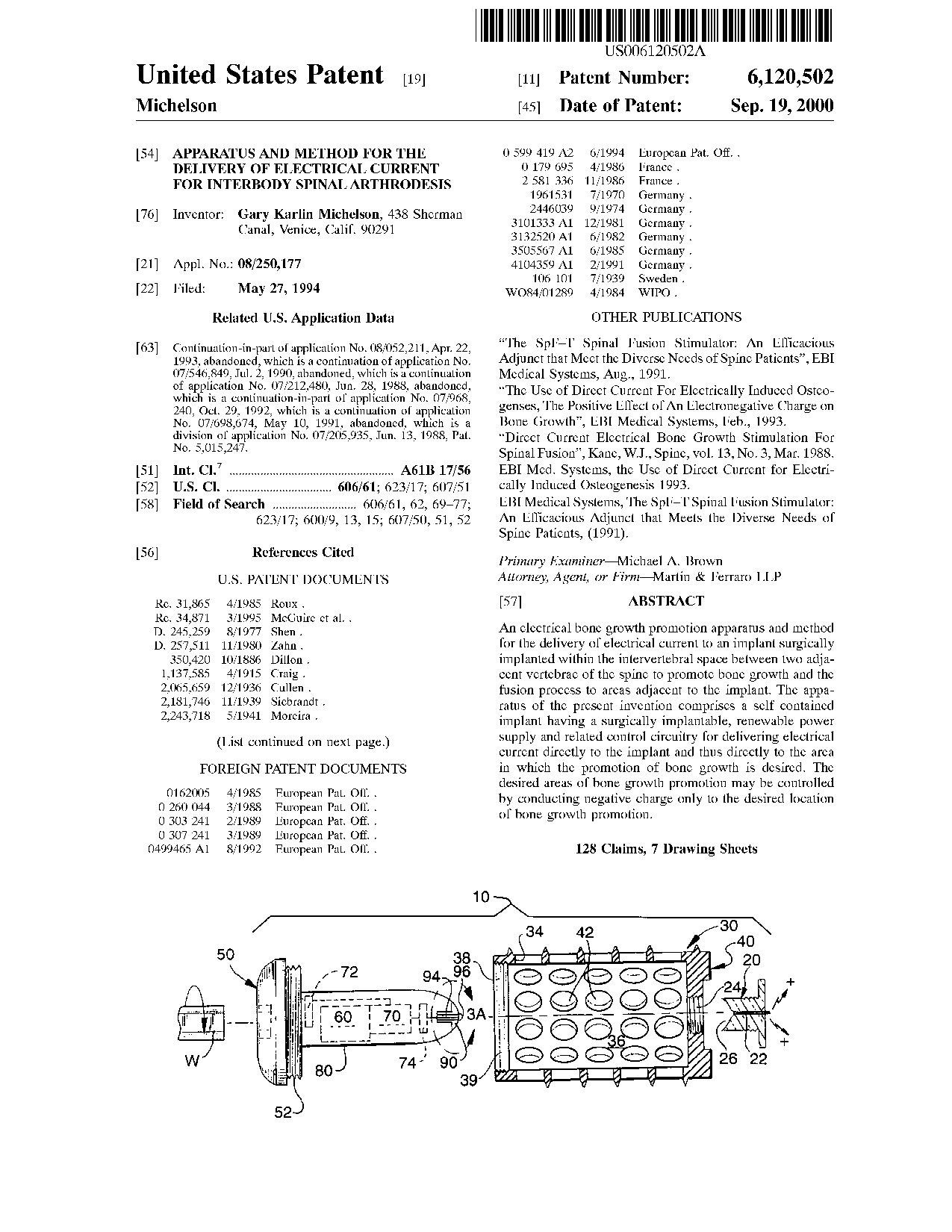 Apparatus and method for the delivery of electrical current for interbody spinal arthrodesis - Patent 6,120,502 Apparatus and method for the delivery of electrical current for interbody spinal arthrodesis - Patent 6,120,502
|
An electrical bone growth promotion apparatus and method for the delivery of electrical current to an implant surgically implanted within the intervertebral space between two adjacent vertebrae of the spine to promote bone growth and the fusion process to areas adjacent to the implant. The apparatus of the present invention comprises a self contained implant having a surgically implantable, renewable power supply and related control circuitry for delivering electrical current directly to the implant and thus directly to the area in which the promotion of bone growth is desired. The desired areas of bone growth promotion may be controlled by conducting negative charge only to the desired location of bone growth promotion.
|
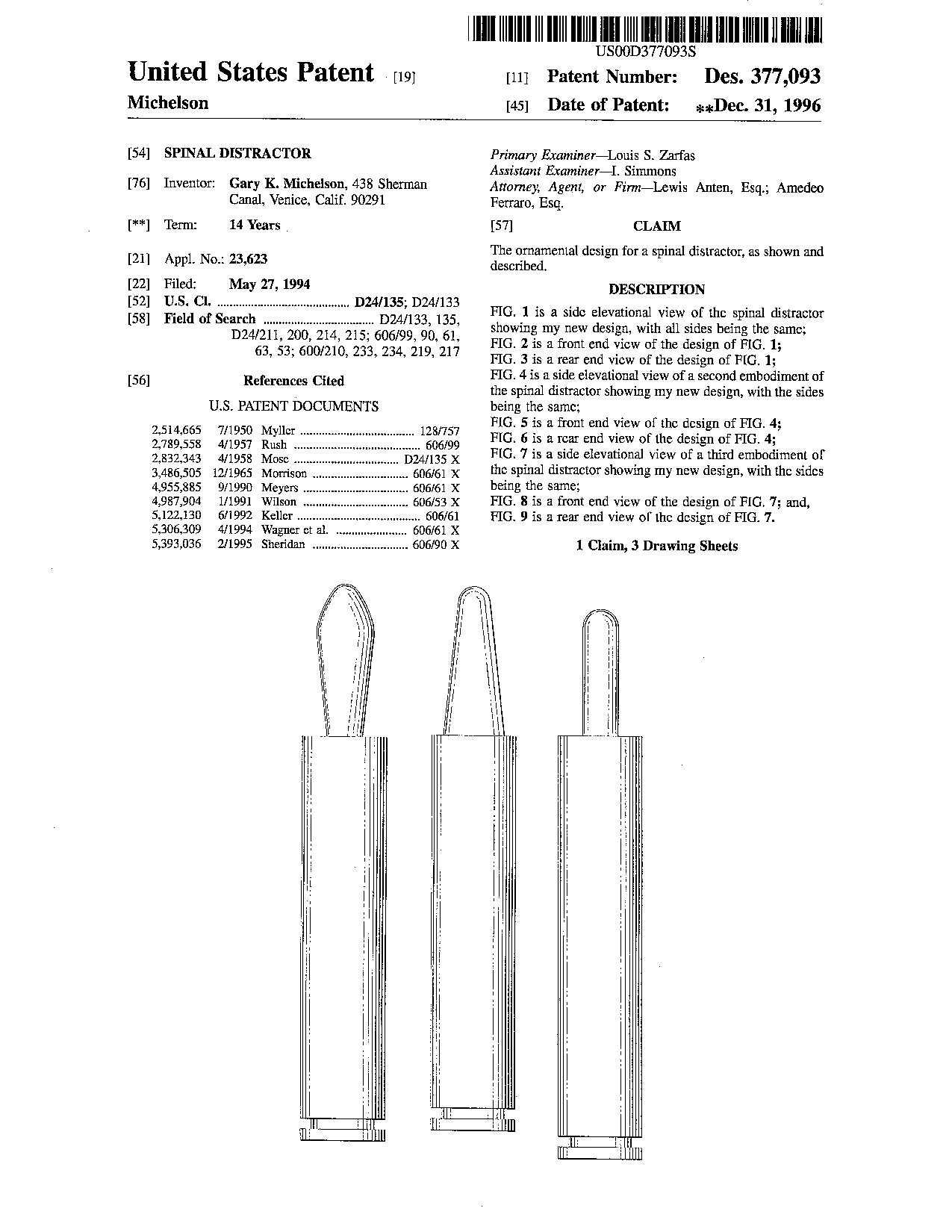 Spinal distractor - Patent D377,093 Spinal distractor - Patent D377,093
|